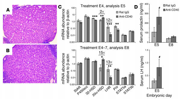
Figure 2.
Postreceptor prolactin signaling defects and luteal insufficiency following systemic CD40 ligation. (A and B) Histological appearance of corpora lutea from mice on E8 following daily E4–7 injections of rat IgG (A) or anti-CD40 antibodies (FGK45) (B). Paraffin sections stained with H&E are shown. (C) Whole-ovary mRNA expression levels determined by real-time RT-PCR. Ovaries were collected from mice that either had been treated with control rat IgG or anti-CD40 antibodies (FGK45) once on E4 and sacrificed 24 hours later on E5 (n = 4 mice per group; upper panel), or had been treated daily on E4–7 and sacrificed on E8 (n = 3 mice per group; lower panel). Data represent mean ± SD. On both E5 and E8, 20α-HSD and LHR mRNA levels were increased and decreased, respectively (fold changes are indicated by brackets), whereas 3β-HSD and prolactin receptor (Prlr) mRNA levels were decreased on E5 only. *P < 0.02; **P < 0.005; ***P < 0.001. (D) Serum prolactin and LH concentrations. Prolactin and LH concentrations in FGK45-treated mice on E5 were significantly elevated compared with those in control mice (#P < 0.05; data represent mean ± SEM of 12 mice per group), but there was no change in prolactin concentrations on E8 (n = 6 mice per group). The limit of detection for LH was 0.2 ng/ml.

