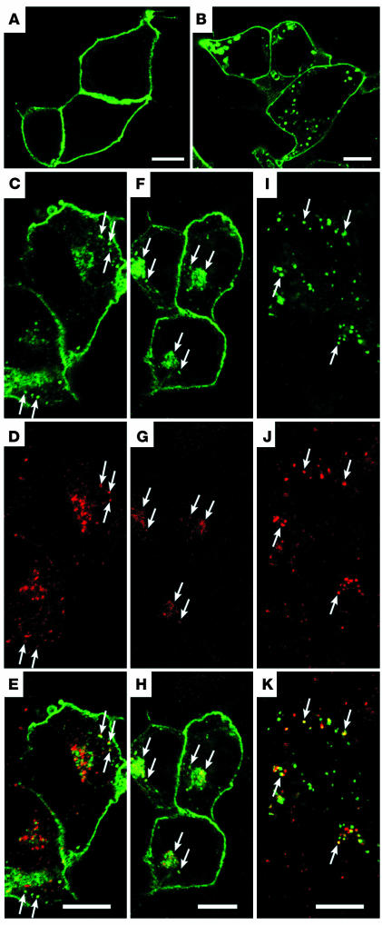
Figure 1.
Confocal images of WT and mutant rhodopsins expressed in HEK cells. (A and B) Fixed HEK cells transfected with WT (A) or R135L (B) rhodopsin were incubated with anti–rhodopsin mAb B6-30 and detected by Alexa 488 secondary antibodies. (C–K) HEK cells transfected with R135L were incubated with Alexa 594–Tf for 5 minutes to label early endosomes (C–E); incubated with Alexa 594–Tf for 2 minutes followed by a 28-minute chase to label recycling endosomes (F–H); or incubated with rhodamine-labeled dextran for 2 hours to label late endosomes/lysosomes (I–K). Cells were then fixed and permeabilized for rhodopsin immunostaining (green). Rhodopsin immunoreactivity colocalized with the internalized Tf or dextran is marked by arrows, and merge images are shown in E, H, and K. Scale bars: 10 μm.

