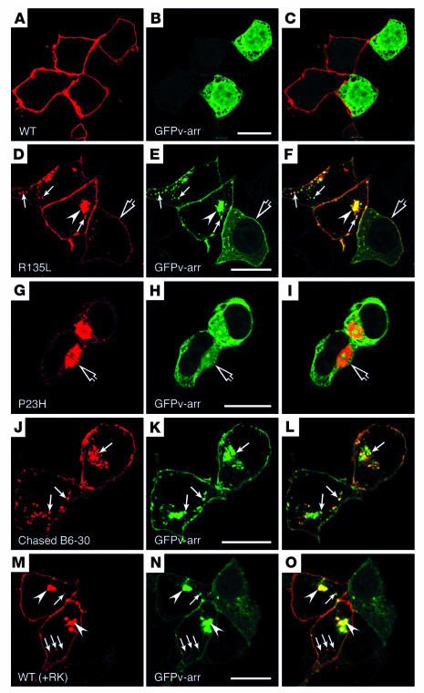
Figure 3.
Subcellular distribution and trafficking of rhodopsin in HEK cells cotransfected with GFPv-arr. (A–I) Cells double transfected with GFPv-arr and either WT rhodopsin (A–C), R135L (D–F), or P23H (G–I) were immunolabeled for rhodopsin (red), and GFPv-arr was directly visualized by its GFP fluorescence. In D–F, some cells expressed low levels of R135L and contained both surface and cytosolic GFP signals (open arrows); the vesicular structures near the cell periphery (arrows) and the perinuclear structure (arrowheads) contain both the R135L and the GFPv-arr. In G–I, the P23H mutant–induced aggresomes (open arrows) are not enriched for GFPv-arr. (J–L) Live, R135L/GFPv-arr–transfected cells were incubated with mAb B6-30 at 4–C, followed by a 1-hour, 37–C chase. The cells were fixed and permeabilized, and the internalized rhodopsin was detected by the labeling of Alexa 594–anti-mouse Ab (red). Internalized surface rhodopsins appeared on GFPv-arr+ vesicle and perinuclear compartments (arrows). (M–O) Cells triple-transfected with WT-rhodopsin, RK, and GFPv-arr were fixed, permeabilized, and either immunolabeled with mAb B6-30 followed by Alexa 594–anti-mouse Ab (M) or directly visualized by GFP signals (N). Phosphorylated WT rhodopsin was colocalized with GFPv-arr on the vesicles underneath PM (arrows) and the perinuclear compartments (arrowheads). A GFPv-arr–singly-transfected neighboring cell exhibited cytosolic green fluorescence. Scale bars: 20 μm.

