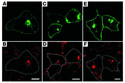
Figure 5.
Aberrant endosomal organization caused by R135L/GFPv-arr. Fixed, transfected cells were incubated with mAb’s that recognized early endosome marker EEA1 (A and B), early/recycling endosome marker TfR (C and D), and late endosome/lysosome marker lysosomal-associated membrane protein 1 (LAMP1) (E and F), followed by Alexa 594–conjugated anti-mouse Ab. GFP signals were used to directly visualize the GFPv-arr. The transfected cells are encircled in B, D, and F to show the cell margins. Although EEA1, TfR, and LAMP1 signals were dispersed in nontransfected cells, their signals were predominantly concentrated in the GFP+ perinuclear structures in the R135L/GFPv-arr–transfected cells. Scale bars: 10 μm.

