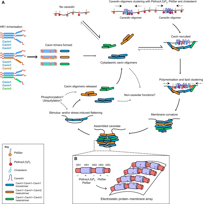
Fig. 3.
A model for cavin function in caveolar formation. (A) The cavin life cycle involves initial trimerisation through the HR1 domain and subsequent assembly into cytoplasmic cavin oligomers. These can bind negatively charged membranes but only become stably associated with the plasma membrane in the presence of caveolins. Caveolins are believed to cluster with cholesterol, PtdIns(4,5)P2 and PtdSer (shown at the top); subsequent recruitment of cavins will further increase the local concentration of the negatively charged lipids in particular. This can then nucleate membrane curvature and formation of the distinctive caveola bud. Certain stimuli, such as membrane stretching or cholesterol depletion, can lead to flattening of caveolae and the release of cavins back into the cytoplasmic pool. Cavins are extensively modified by posttranslational modifications, but the negative or positive consequences for caveolar formation are still unknown. (B) An expanded view of the membrane surface, showing a speculative model for cavin–cavin interactions through an array of electrostatic interactions involving both membrane lipids and domains of opposite charge in adjoining protomers.

