Fig. 6.
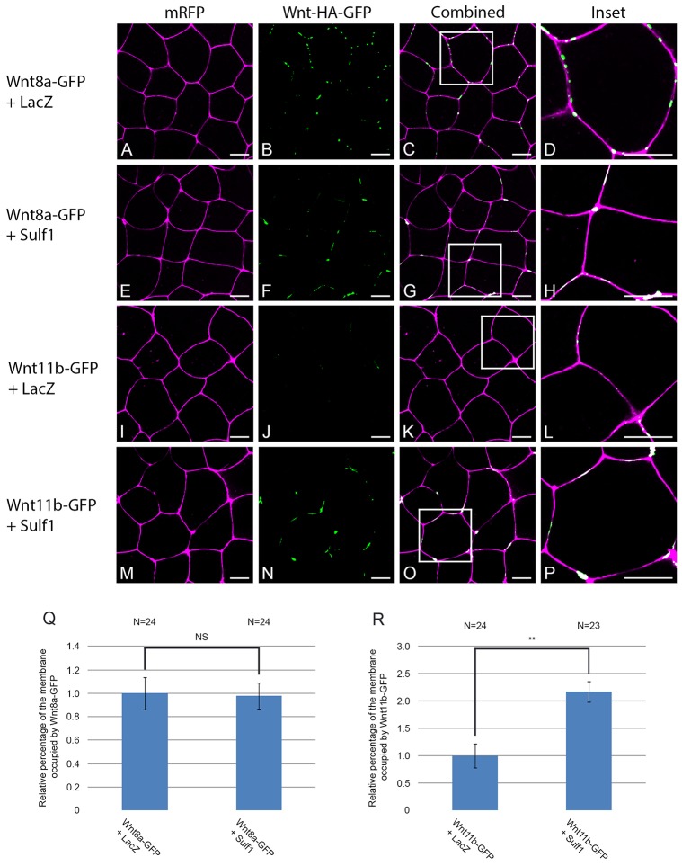
Sulf1 enhances the accumulation of Wnt11b–GFP on the cell membrane. (A–P) Xenopus laevis embryos were microinjected bilaterally with mRNA encoding mRFP (500 pg) into the animal hemisphere at the two-cell stage. In addition, embryos were injected with mRNA encoding LacZ (4 ng), Sulf1 (4 ng), Wnt8a–GFP (500 pg), Wnt11b–GFP (1 ng) or a mixture of the four. (A–D) Control animal explants overexpressing LacZ and Wnt8a–GFP. (E–H) Animal explants overexpressing Sulf1 and Wnt8a–GFP. (I–L) Control animal explants overexpressing LacZ and Wnt11b–GFP. (M–P) Animal explants overexpressing Sulf1 and Wnt11b–GFP. The white boxes in C, G, K and O mark the areas that are enlarged in D, H, L and P, respectively. mRFP is shown in magenta, Wnt8a– or Wnt11b–GFP is shown in green. Scale bars: 20 μm. (Q,R) Graphs quantifying the relative levels of (Q) Wnt8a–GFP and (R) Wnt11b–GFP on the cell membrane. Data was quantified using a programme written in Matlab, results are mean±s.e.m. **P<0.01; NS, not significant (Mann–Whitney U test). N, number of embryos.

