Fig. 8.
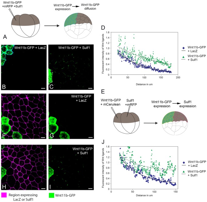
Sulf1 enhances both the secretion and the amount of Wnt11b–GFP diffusing in animal explants. (A) Diagram depicting the assay used to measure Wnt11b–GFP secretion and diffusion through a control background. (B,C) mRNA encoding either (B) mCerulean (600 pg), LacZ (4 ng) and Wnt11b-GFP (2 ng) or (C) mRFP (600 pg), Sulf1 (4 ng) and Wnt11b–GFP (2 ng) was injected into the animal hemisphere of one blastomere at the four-cell stage. (D) The range of diffusion of Wnt11b–GFP through a control background was quantified using Fiji Image J. (E) Diagram depicting the assay used to measure Wnt11b–GFP diffusion through a background expressing Sulf1. (F–I) mRNA encoding mCerulean (600 pg) and Wnt11b–GFP (2 ng) was injected into the animal hemisphere of one blastomere at the four-cell stage. An adjacent blastomere was injected with mRNA encoding (F,G) mRFP (600 pg) and LacZ (4 ng) or (H,I) mRFP (600 pg) and Sulf1 (4 ng). (J) The range of Wnt11b–GFP through a background expressing either LacZ or Sulf1 was quantified using Fiji Image J. Scale bars: 20 μm.

