Figure 4.
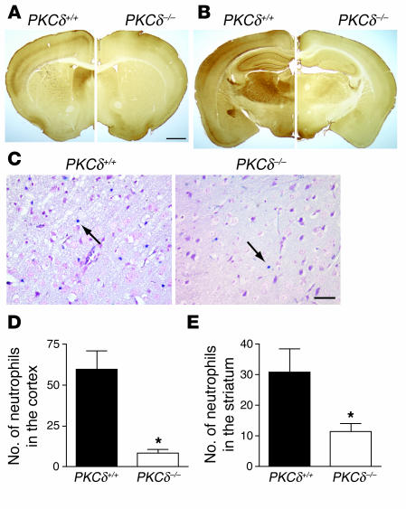
Expression pattern of PKCδ in the brain and extravascular neutrophils after transient MCA occlusion. (A and B) Immunocytochemical localizations of PKCδ in the mouse brain are shown in coronal sections at bregma 0.38 mm (A) and a more posterior section at bregma –1.58 mm (B) for PKCδ+/+ (left) and PKCδ –/ – (right) mice. (C) Representative sections from PKCδ+/+ and PKCδ –/ – mice showing reduced neutrophil accumulation within infarcted tissue in the cortex of PKCδ –/ – mice after 1 hour of MCAO and 24 hours of reperfusion. Blue infiltrated neutrophils were identified in the ischemic cortex by staining for esterase activity with dichloroacetate (arrows). (D and E) Number of extravascular neutrophils in the ischemic cortex and striatum of PKCδ+/+ (n = 7) and PKCδ –/ – mice (n = 6) after transient MCAO. No esterase staining was seen in sections from nonischemic animals (data not shown). The scale bars in A and C correspond to 1 mm and 25 & μ;m, respectively. *P < 0.05 compared with WT littermates.

