Figure 1.
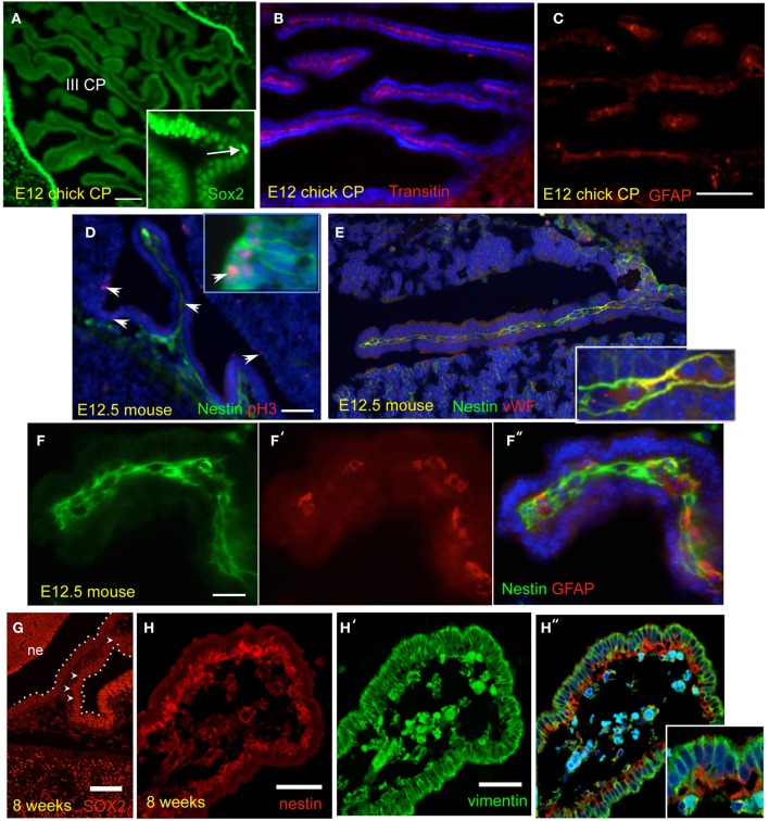
Neural progenitor markers are expressed in the developing choroid plexus (CP), in different species. Unless otherwise indicated, micrographs show the CP of lateral ventricles. (A–C) E12 Chick CP stained for Sox2, transitin, and GFAP. III CP: third ventricle CP. (D–F) E12.5 mouse CP stained for pH3 (phosphorylated-histone 3), nestin, von Willebrand factor, and GFAP, either alone or in combination. Arrowheads in (D) point at proliferating cells in the CP and in the neuropepithelium (insert); only the CP is shown in (F). (G,H) Human CP at 8 weeks of gestation (CS23) stained for Sox2, nestin, and vimentin. The CP is outlined and some of the brighter SOX2-positve cells in the CP are indicated by arrowheads; note the gradient of SOX2 staining in the CP; ne, neuroepithelium. Nuclei are counterstained with Hoechst dye (blue). Scale bars are: (A) = 50 μm; (B,C) (same magnification) = 100 μm; (D,E) (same magnification) = 100 μm; (F) = 100 μm; (G) = 100 μm; (H) = 50 μm.

