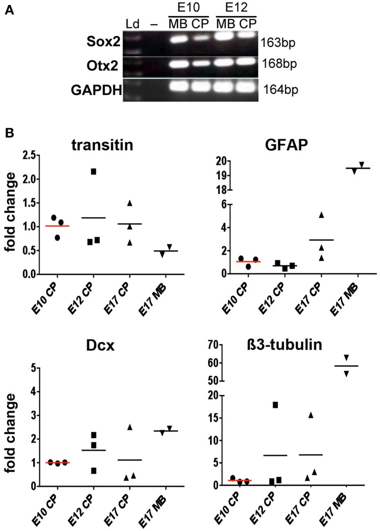
Figure 2.
Expression of neural transcripts in the chick lateral ventricle choroid plexus (CP) at E10, E12, and E17 detected by RT-PCR and RT-qPCR. (A) Detection of Sox2 and Otx2 by RT-PCR in the CP and midbrain (MB, positive control). (B) Detection of transitin, GFAP, Dcx, and β3-tubulin by RT-qPCR. Fold changes in individual CPs or MB normalized against GAPDH are shown. Note that transitin, GFAP, Dcx, and β3-tubulin transcripts are detected in the CP at all developmental stages examined; no statistically significant differences in expression levels are observed.

