Figure 3.
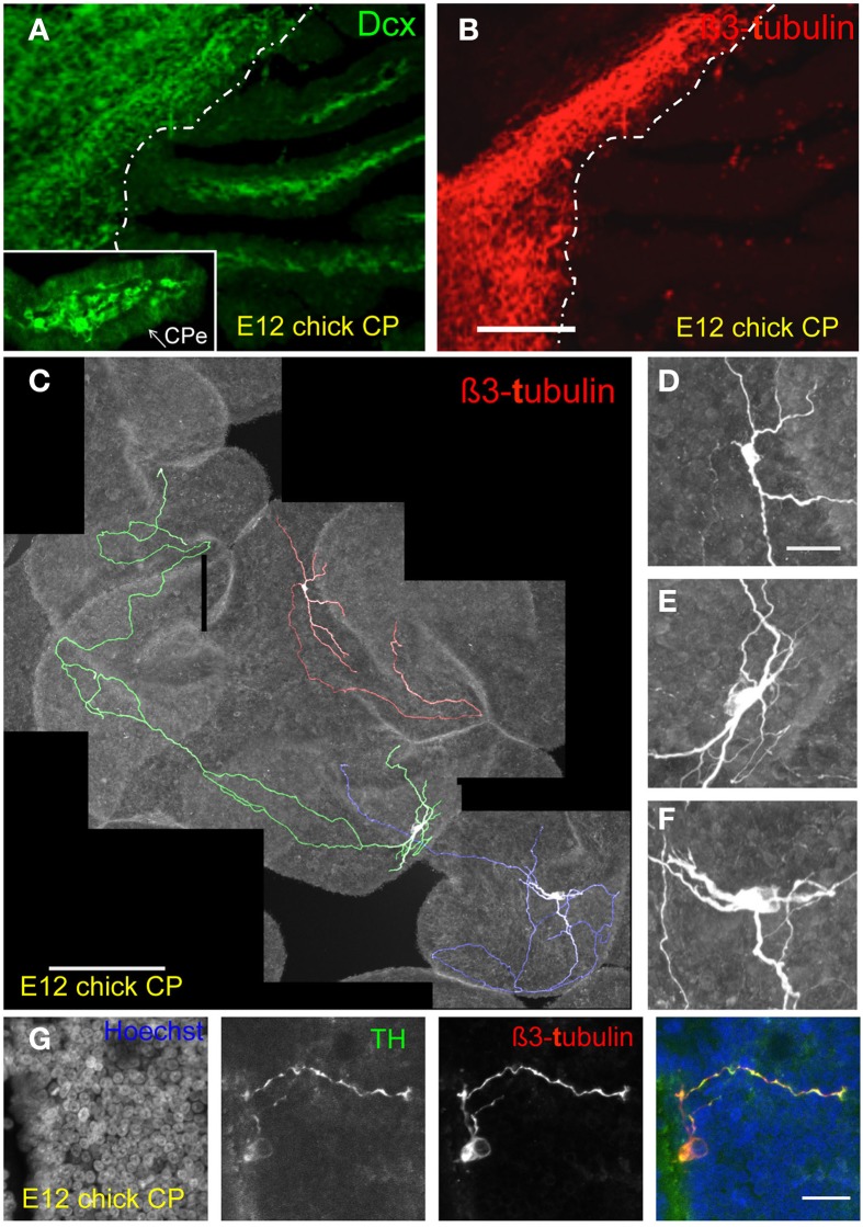
Identification of neuroblasts and neurons in the developing chick CP. (A,B) E12 lateral CP section stained for doublecortin (Dcx) and ß3-tubulin in E12 chick CP. Note Dcx reactivity in the CP stroma (insert in A) as well as in the brain; only some punctuated staining is observed in the CP with the anti-ß3-tubulin antibody whereas the brain tissue is strongly labeled. The approximate boundary between brain and CP is indicated by the dotted line. CPe, CP epithelium. (C–F) Examples of extensive branching (individual neurons in (C) are shown in different colors for ease of visualization) and neuronal morphologies detected by ß3-tubulin staining. (G) Staining of the E12 CP for tyrosine-hydroxylase (TH), ß3-tubulin and Hoechst dye (nuclei) and merged image; note the presence of a positive TH neuron in proximity of the CP epithelium. Scale bars: (A,B) (same magnification) = 200 μm; (C) = 200 μm; (D–F) (same magnification) = 30 μm; (G) = 20 μm.

