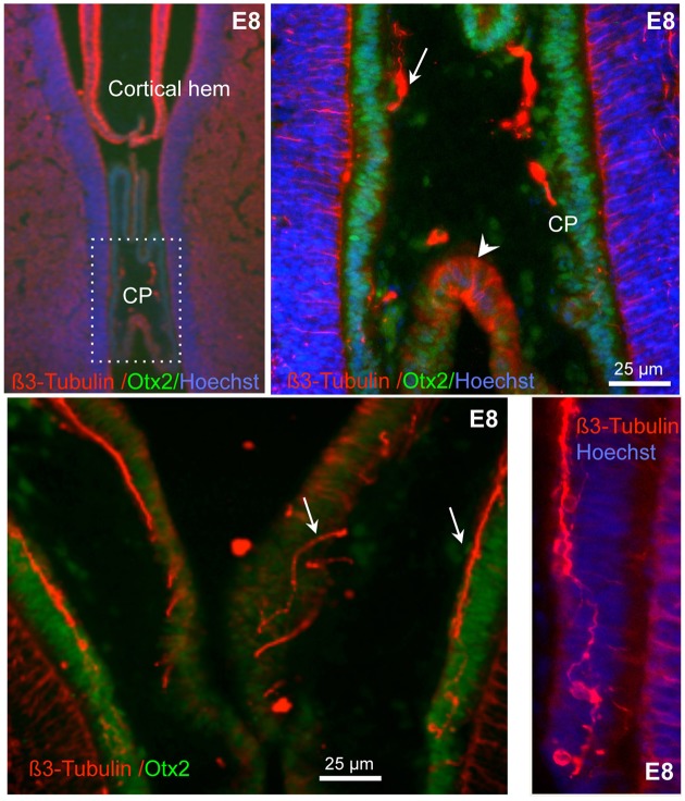
Figure 4.
Expression of β3-tubulin in E8 chick CP. Sections double-labeled for ß3-tubulin and Otx2, a marker of the CP epithelium. High magnification images are shown in the right panels. Note the presence of nerves and neurons (some indicated by arrows) in the CP as well as of cells spanning across the CP epithelium (arrowhead). Nuclei are counterstained with Hoechst (blue). The bottom right panel is at the same magnification as the one at its left.

