Figure 7.
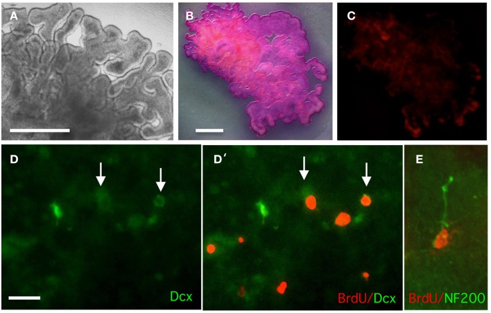
Analysis of survival, proliferation, and doublecortin (Dcx) expression in organotypic cultures of E12 lateral CP. (A) Phase image of CP after 7 days in culture. (B) MTT staining of live CP after 7 days in culture; extensive staining is indicative cell viability. (C) Propidium iodide staining of live CP after 7 days in culture to detect dead cells; only very limited staining is observed. (D,D') CP organotypic culture treated with BrdU for 3 days and double-stained for BrdU and doublecortin (Dcx). Some double-labeled cells are observed. (E) Detection of NF200 and BrdU in an organotypic culture. Scale bars: (A–C) = 200 μm (B and C are at the same magnification); (D,E) = 50 μm.

