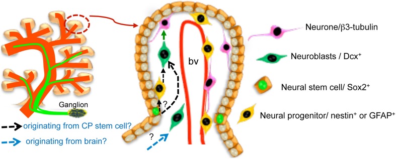
Figure 8.
Schematic representation of the CP neural components (innervation from ganglia outside the CP and neurons within the CP) and of the model proposing the existence of a neurovascular niche within the developing chick CP. It is not currently known whether nestin-like-positive cells reflect a transition from high-Sox2/vimentin-positive cells to neuroblasts (black dotted arrows) nor whether some of the CP neurons are born from neuroblasts migrating into the CP from the brain rather than having been generated within it (blue dotted arrow). Please note that only the subset of highly Sox2-positive putative progenitors in the CP epithelium is indicated in the cartoon. The two sources are not mutually exclusive. bv, blood vessel.

