Figure 3.

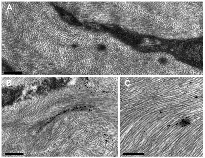
Electron micrographs of EFMBs in WT mouse. (A) Shows a keratocyte with 3 EFMBs nearby. Panels (B & C) show fibrillin microfibrils labeled with immuno-gold particles and enhanced with silver. (Scale bars; (A) 500 nm, (B) 750 nm, (C) 500nm)

Electron micrographs of EFMBs in WT mouse. (A) Shows a keratocyte with 3 EFMBs nearby. Panels (B & C) show fibrillin microfibrils labeled with immuno-gold particles and enhanced with silver. (Scale bars; (A) 500 nm, (B) 750 nm, (C) 500nm)