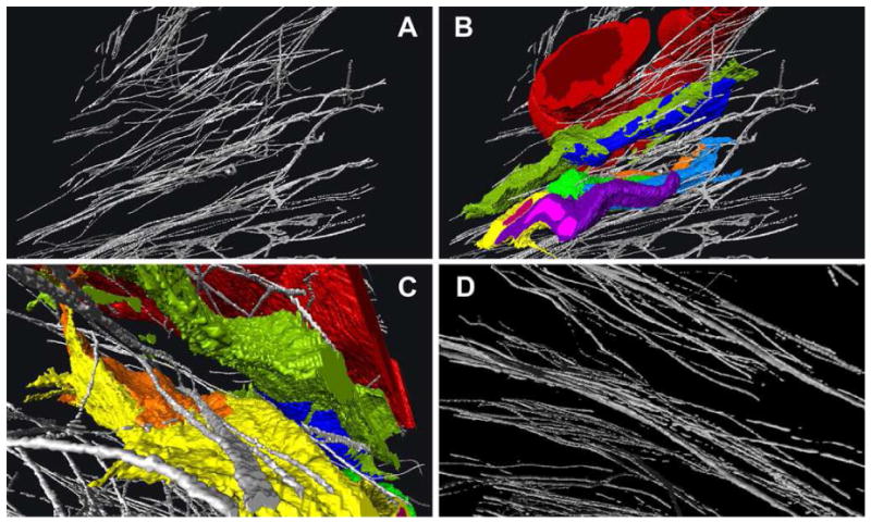
Figure 4.

3D reconstructions from serial SEM block-face images. Panel A shows EFMBs in the limbus, and panel B the same area with blood vessel (red), keratocytes (green, blue, yellow) and neutrophil (purple) in a mouse cornea 6 hours after epithelial wounding. Panel C is from the same 3D reconstruction as A and B but from a different angle and zoomed in to show more detail. From this perspective some EFMBs appear to lie within a groove in the surface of a keratocyte. Panel D is a reconstructed image showing EFMBs in the paracentral cornea with more defined layering as compared to the limbus. Panels A-C are views from an oblique angular perspective while the line of projection in panel D is nearly parallel with the corneal surface. The average EFMBs were 100-200nm in diameter when measured in cross-section block face images.
