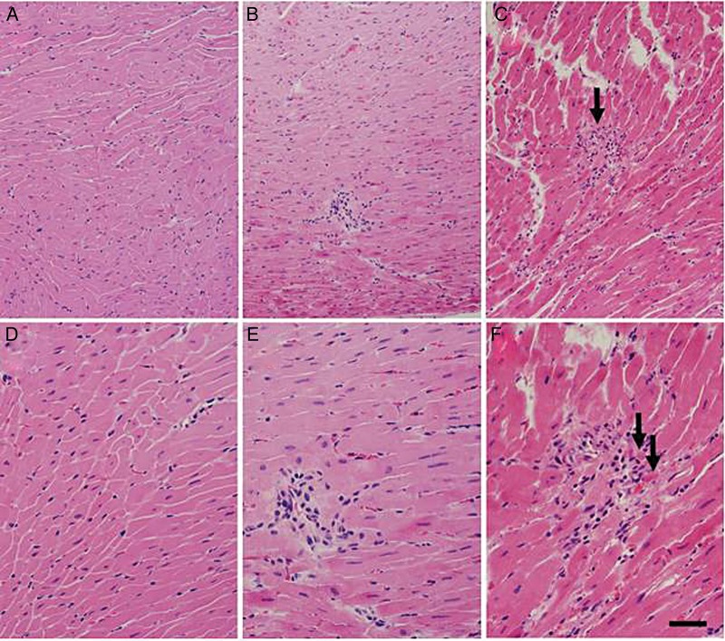
Fig. 6.
Histological findings in irradiated rat hearts. H&E stains of hearts taken at two magnifications from unirradiated (A and D) and irradiated (B, C, E and F) rats reveal scattered foci of lymphocytic inflammation in a subpopulation of irradiated rats. One irradiated rat showed cardiac myocyte necrosis (C and F, arrows) associated with lymphocytic inflammation. Bars = 80 μm (A, B and C) and 40 μm (D, E and F).

