Figure 4.
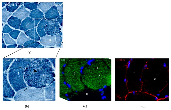
Intermyofibrillar network changes in muscle fibers lacking sarcolemmal nNOS. Enlarged type I fiber (#) with decreased sarcolemmal nNOS protein shows alterations in the intermyofibrillar network demonstrated by irregular NADH-TR staining ((a) and (b): arrow head). Note also a subsarcolemmal accumulation of cellular material or mitochondria seen as dark areas below the fiber membrane ((b): dark arrow). Type II fibers appear normal. NADH-TR (mitochondria) is blue, MHC-I is green, and nNOS is red. # marks identical fibers on serial sections. Scale bar: 50 μm.

