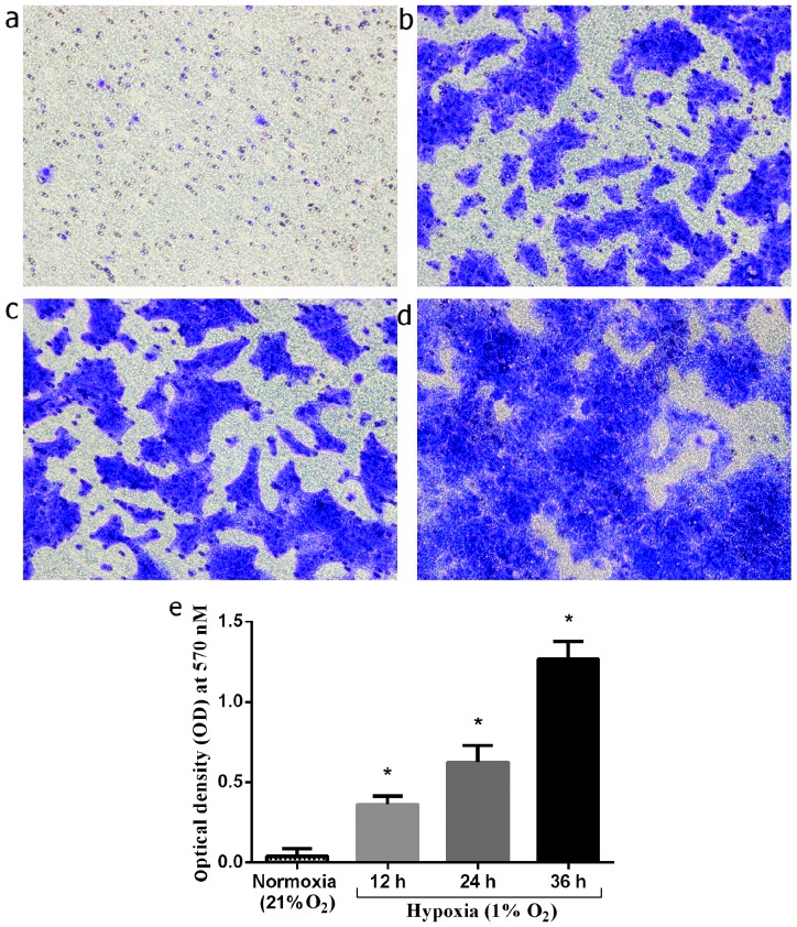
Figure 7.
Cell invasiveness investigated by Matrigel co-culture chambers. The invasive cells were fixed and stained with Crystal violet. (a) Invasion of keloid keratinocytes under normoxic conditions (21% O2). (b–d) Invasion of keloid keratinocytes cultured under hypoxia conditions (1% O2) for 12, 24 and 36 h, respectively. (e) Columns indicate the optical density (OD) of Crystal violet eluted from the invading keloid keratinocytes under normoxic and hypoxic culture conditions (12, 24 and 36 h). *P<0.05 vs. keratinocytes under normoxic conditions (21% O2). Bars represent the means ± SD of 3 independent experiments.

