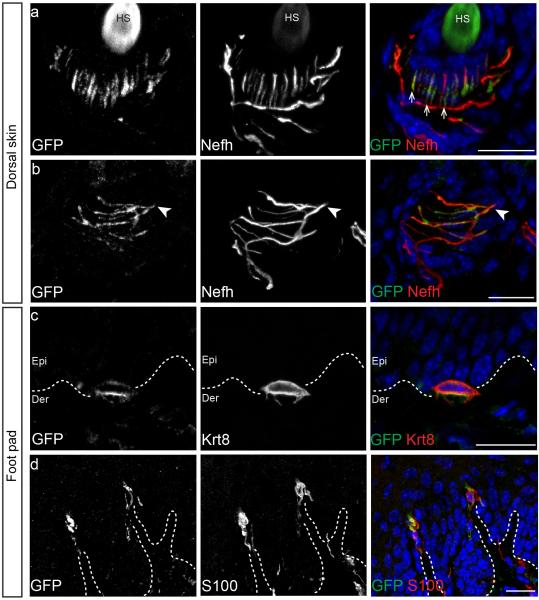
Figure 1. Piezo2 is localized at the nerve terminals of sensory neurons that innervate the skin.
a and b, representative images of immunostaining of GFP, Nefh and DAPI (blue) in the hair follicle of the Piezo2-GFP dorsal skin. GFP staining indicates localization of the Piezo2-GFP fusion protein and Nefh marks myelinated neurons. c, representative image of immunostaining of GFP, Krt8, a specific marker of Merkel cells, and DAPI (blue) in the Piezo2-GFP glabrous skin. d, representative images of immunostaining of GFP and S100, a marker of Schwann cells, in glabrous skin. Arrows mark lanceolate endings (a) and arrowheads mark circumferential fibers (b). Dashed lines demarcate epidermal-dermal junction (c and d). HS, hair shaft; Epi, epidermis; Der, dermis. Scale bars (a-d) 20μm.

