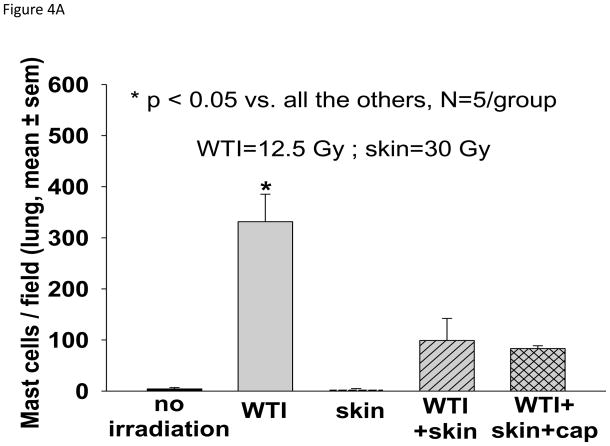
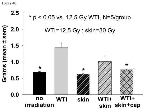
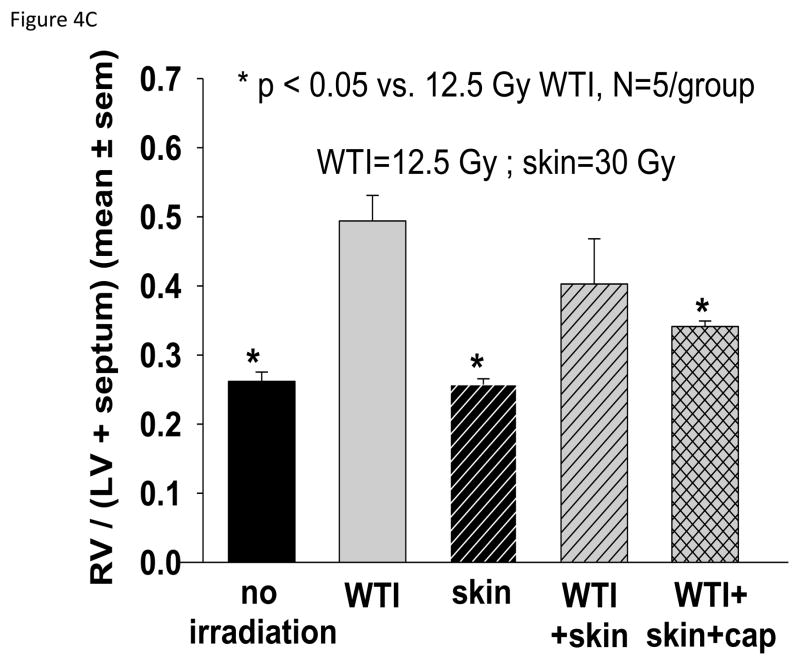
FIGURE 4.
Histology and structure of the lung and heart at 42 days after 12.5 Gy irradiation (for details see Materials and Methods). Lungs and hearts were harvested from 5 groups (treatments i–v, see Materials and Methods). The lungs were weighed, fixed, sectioned and stained with anti-tryptase antibody. The hearts were dissected and the right ventricle as well as the left ventricle and septum were weighed separately. 4A. Representations of the mean±SEM of the number of mast cells/field in 5 experimental groups as labeled. N= 5 rats/group. Note the large increase in mast cells from lung sections of rats irradiated to the thorax only. B. Mean±SEM of wet weight of the lungs in each group. The lung weight doubled at 42 days after 12.5 Gy WTI. C. Depicts the ratio of the right ventricle:left ventricle+septum. Note the right ventricular hypertrophy in rats that received WTI, was mitigated by captopril.


