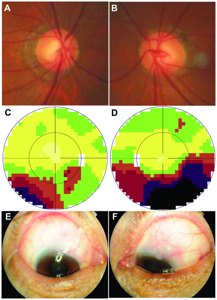
Figure 2.
Images were obtained from one primary open-angle glaucoma (POAG) patient (096003) in the family. (A) Fundus image of the right eye, the C/D ratio is ~0.6, and the myopia arc is visible. (B) Fundus photograph taken from the left eye, the C/D ratio is ~0.7, and the myopia arc is also visible. (C) The map of the visual field (Octopus) of the right eye. The lower part of the visual field was defective, shown as brown, red and purple on the map. (D) The map of the visual field of the left eye. The lower half of the visual field was defective, shown as brown, red, purple and black on the map. The defect was more severe than that of the right eye. (E and F) Functional conjunctival blebs were observed in a follow-up examination following trabecule ctomy. Conjunctival hyperemia was not apparent.

