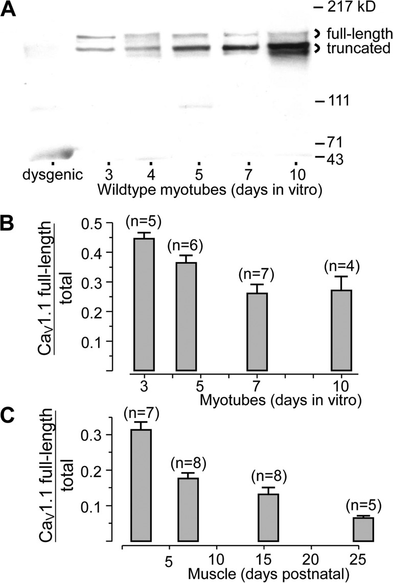
Figure 2.
Developmental changes in expression of full-length and truncated CaV1.1. (A) Western blots of membranes isolated from wild-type myotubes grown in culture for the indicated number of days (denoting plating as “Day 1”) and probed with the IIF7 monoclonal antibody, which recognizes both the full-length and truncated species. The leftmost lane illustrates membranes from dysgenic (CaV1.1-null) myotubes. Based on scanning densitometry, full-length CaV1.1 as a fraction of total (full-length plus truncated) is plotted as a function of developmental age in vitro (B) and in vivo (C). Error bars indicate SEM, and numbers of cultures or mice are indicated in parentheses. In B, myotubes from 4–5 d were grouped and plotted at day 4.5. In C, mice of 1–3, 5–9, 13–18, and 21–30 d, respectively, were grouped and plotted at time points corresponding to the average age of each group.

