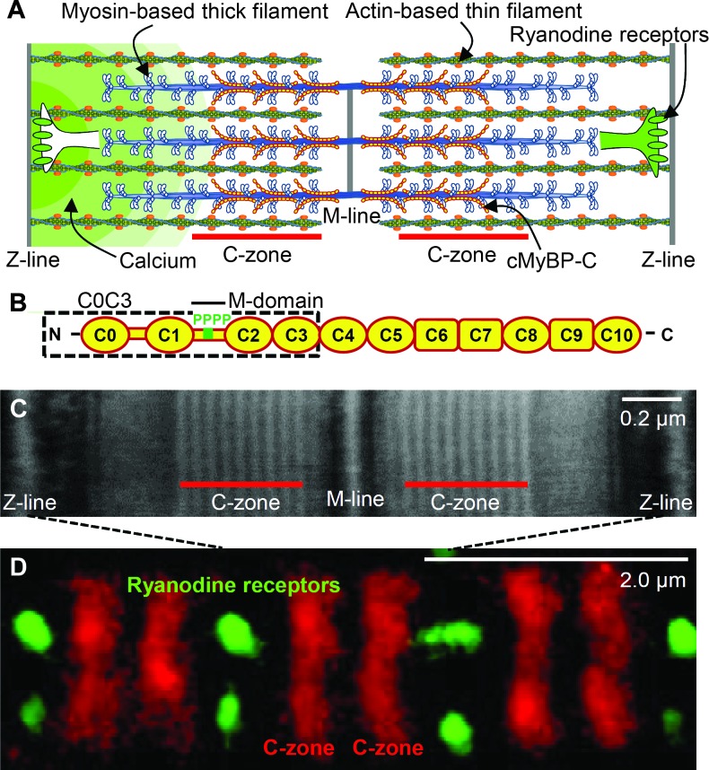
Fig. 1. Sarcomeric organization and MyBP-C.
(A) Cardiac muscle sarcomere with interdigitating thick and thin filaments. MyBP-C localized to the C-zone, whereas the ryanodine receptors are localized in puncta (CRUs) along the Z-lines, forming the boundaries of each sarcomere. (B) Schematic diagram of cardiac MyBP-C’s Ig-like (oval) and fibronectin-like (rectangular) domains with four phosphorylation sites (P) in the M-domain and C0C3 fragment (dashed box) used in the 3D EM and in vitro motility experiments. (C) Negatively stained EM image of a sarcomere within a mouse ventricular myocyte labeled with antibodies to MyBP-C. (D) Two-color dSTORM super-resolution image of sarcomeres as in (C), labeled with Alexa Fluor–conjugated antibodies to MyBP-C and ryanodine receptors.

