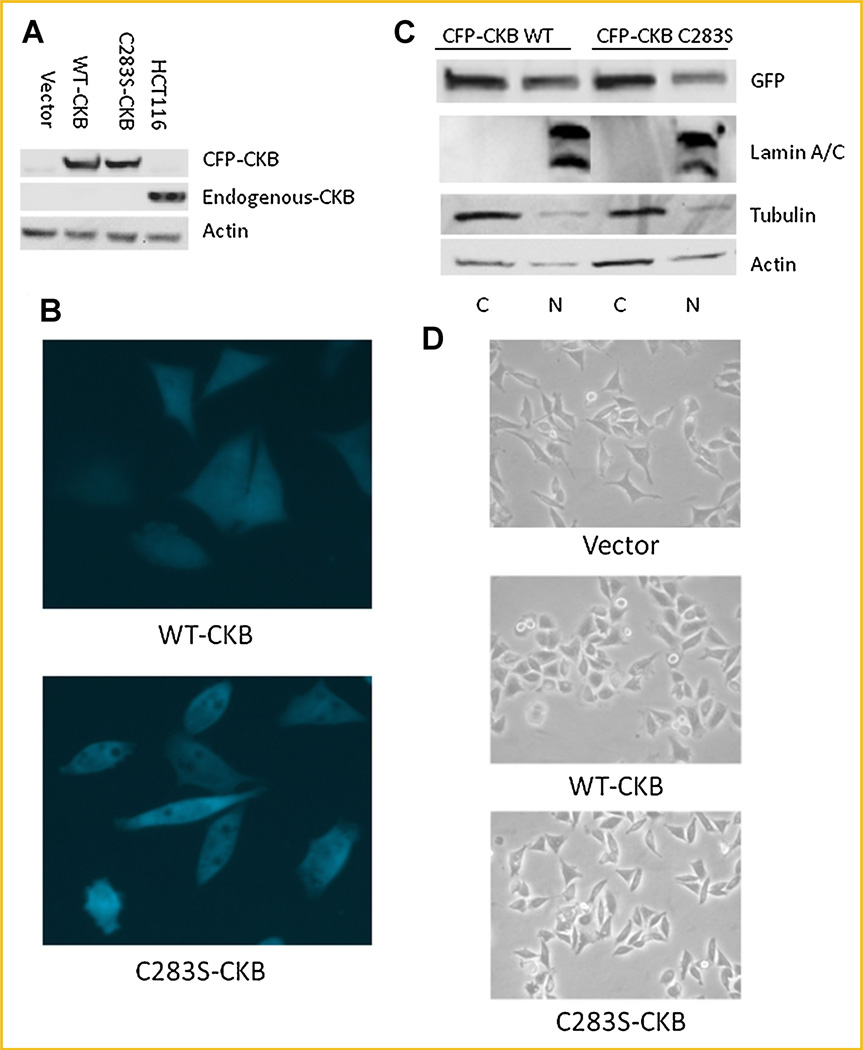
Fig. 2.
WT- and C283S-CKB have similar localization in stably transfected Caco-2 cells. A: Cells were lysed and immunoblotted for expression of CKB and actin, with HCT116 whole-cell lysate used as a positive CKB control. B: The relative distributions of CFP-CKB constructs were measured in whole cells by fluorescence microscopy (40×). C: Cells were fractionated into nuclear and cytoplasmic pools. Immunoblotting was performed with a CFP antibody to show the relative distribution of transfected protein. D: Light microscopy (20×) of the three cell lines was used to determine overall cell shape. [Color figure can be viewed in the online issue, which is available at wileyonlinelibrary.com.]

