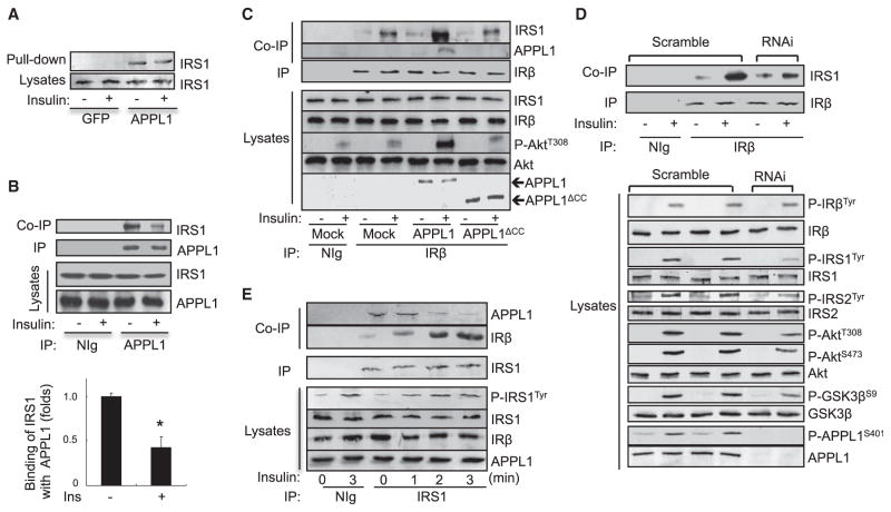
Figure 5. APPL1 Facilitates the Recruitment of IRS1 onto IRβ.
(A) Glutathione agarose beads containing GST, GST-APPL1 fusion proteins were incubated with lysates of C2C12 myotubes treated with or without 10 nM insulin (3 min). Endogenous IRS1 associated with GST-APPL1 fusion proteins was detected with an antibody specific to IRS1.
(B) Serum-starved C2C12 myotubes were treated with or without 10 nM insulin (3 min). Endogenous APPL1 was immunoprecipitated with an antibody to APPL1, and the coimmunoprecipitated IRS1 was detected by western blot analysis using an antibody to IRS1. Data are shown as mean ± SEM from three independent experiments. p values were calculated using the Student’s t test (*p < 0.05).
(C) C2C12 myocytes transfected with or without myc-tagged APPL1 or APPL1ΔCC truncation were serum starved and treated with or without 100 nM insulin (3 min). Endogenous IRβ was immunoprecipitated, and coimmunoprecipitated endogenous IRS1 or myc-tagged APPL1 was detected with specific antibodies as indicated.
(D) Scrambled control and APPL1-suppressed C2C12 myotubes were serum starved and treated with or without 10 nM insulin (3 min). Endogenous IRβ was immunoprecipitated, and coimmunoprecipitated IRS1 was detected by western blot analysis with the antibodies as indicated.
(E) Serum-starved C2C12 myotubes were treated with or without 10 nM insulin for the indicated times. Endogenous IRS1 was immunoprecipitated, and coimmunoprecipitated endogenous IRβ and APPL1 were detected by western blot analysis with the antibodies as indicated.

