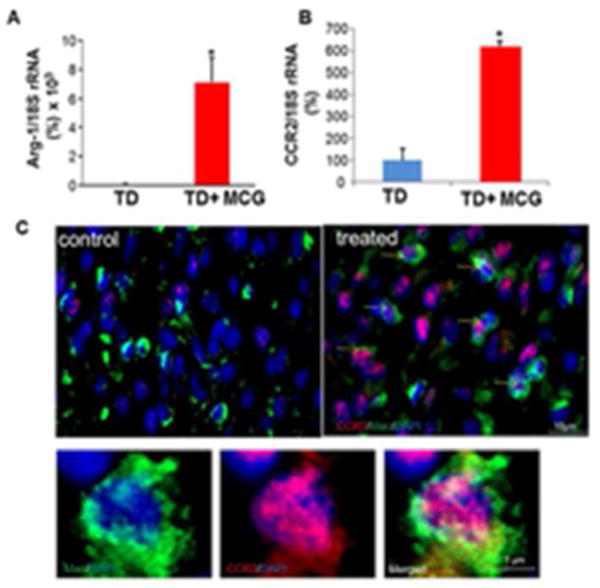
Figure 3. increased CCR2 (M2c macrophage marker) expression in macrophages treated with modified collagen gel (MCG).

(A&B) Real-time PCR was used to measure Arg-1 & CCR2 gene expressions in wound tissue samples at day 7 post- wounding. Gene expression data are presented as % change compared to untreated wound tissues. Data are mean ± SEM; *p < 0.05. (C) Representative images of control and treated wound-edge tissues immunostained using Anti-MAC387 (macrophages, green) and Anti-CCR2 E68 (M2c macrophages, red). The sections were counterstained with DAPI (blue). Scale bar, 10 μm. Insets are zoomed regions in the image, Scale bar, 1 μm.
