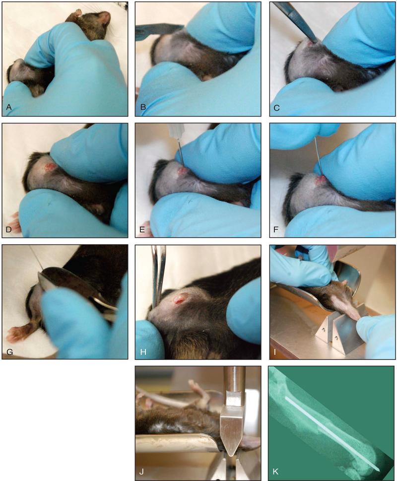
Figure 2. Fracture procedure.
A: The knee is maximally flexed and help using the thumb and index finger of the non-dominant hand. B: A 2-3 mm longitudinal incision is made centered over the knee. C: A subsequent incision is made medial to the patella, which extends into the quadriceps muscle and along the patella tendon to release the tissue. The patella is subluxated laterally. D: Exposure of the distal femur. E: Entry portal to distal femur created using 27-gauge needle in the center of the femoral groove. F: Insertion of femoral pin down the length of the medullary canal. G: This is cut flush with the distal femur. H: The extensor mechanism is placed back to its anatomic location. I, J: Demonstrates creation of closed mid-shaft femur fracture. K: Example of completed fracture.

