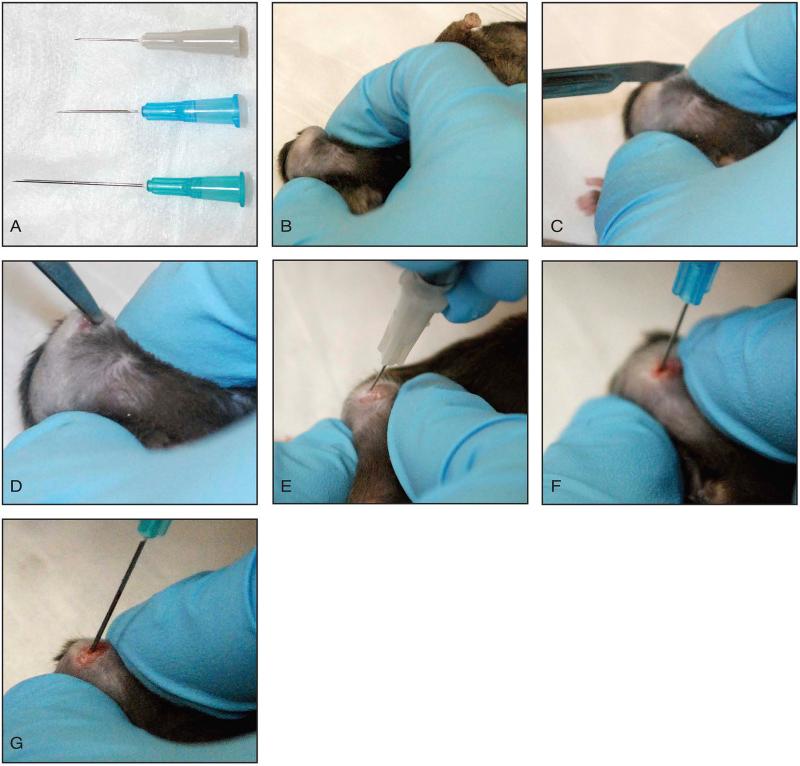
Figure 4. Marrow ablation procedure.
A: Three different needle sizes required for progressive ablation of the tibia. B: The knee is maximally flexed. C: A 2-3 mm longitudinal incision is made centered over the knee. D: An incision is made medial to the patella, which extends into the quadriceps muscle and along the patella tendon to release the tissue. The patella is subluxated laterally. E, F, G: The proximal tibia is instrumented and progressively larger needles are used to ablate the medullary canal.

