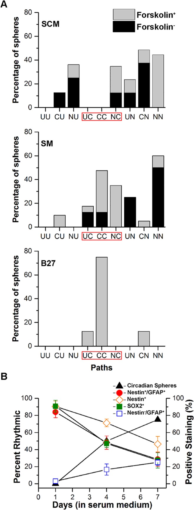
Fig 2. The rhythmic state of spheres during early and late exposure to three culture conditions.

A: Spheres were maintained in either SCM, SM or B27 medium. Spheres were imaged immediately after a forskolin treatment to synchronize circadian clock cells or after no treatment. Shown is the percentage of spheres that began in a particular state (C: circadian, U: ultradian, N: nonrhythmic) during the first 3 days of imaging (early) and their state during the final 3–4 days (late) of imaging sessions. Under differentiating conditions (SM or B27) the three paths to the circadian state (UC, CC, and NC) were most commonly observed. B: The increase in the percentage of spheres showing circadian rhythms is correlated with an increase in the differentiation marker Nestin-/GFAP+ and negatively correlated with the decline in stem cell markers (Nestin+/GFAP+, Nestin+, and SOX2+) during 7 days in SM.
