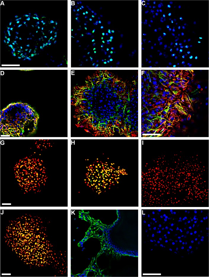
Fig 3. Emergence of circadian rhythms before fully differentiated neurons appear.
Spheres were synchronized by forskolin treatment and fixed after differentiation in SM or B27 medium, mimicking BLI conditions. Hoechst (blue) or propidium iodide (red) were used as nuclear stains. NSPCs were identified as SOX2+ (cyan; A-C: after 1, 4, 7 days in SM), Nestin+/GFAP+ (yellow; D-F: after 1, 4, 7 days in SM; red: GFAP, green: Nestin) or Msi1+ (yellow; H: after 3 days in SM). Additional spheres were fixed after differentiation in medium with serum or B27 supplement to stain for progenitor cells as Dcx+ (Yellow; G, J: after 4 days in SM or B27, respectively). Immature neuronal cells were identified as BetaIII-tubulin+ (Green; K: after 5 days in B27 medium), and mature neuronal cells as NeuN+ (Yellow; I: after 4 days in SM and Green; L: after 4 days in B27 medium). Scale bars = 50 μm, and A-C, E, H, I, and K are at the same magnification.

