
Figure 3. Host susceptibility to DNA viral infection after pretreating with RXR agonists and antagonist.
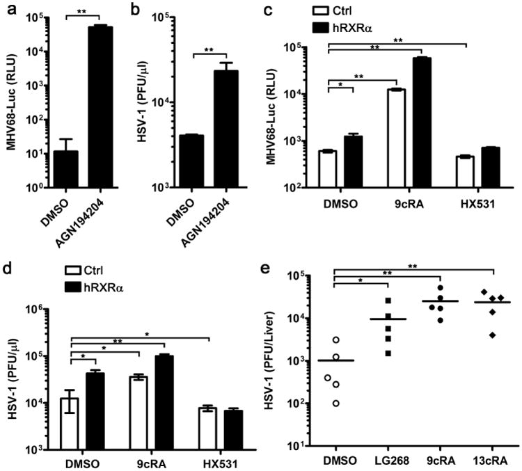
(a) hRXRα-overexpressing RAW264.7 cells were treated with DMSO or AGN19420 (100 nM) for 16 h and subsequently infected by luciferase-expressing MHV68 (MHV68-luc; MOI = 0.5). Cells were lysed in passive lysis buffer at 24 h.p.i and the luciferase activity was quantified by luminescence assay. (b) hRXRα-overexpressing RAW264.7 cells pretreated with DMSO or AGN19424 (100 nM) and then these cells were infected by HSV-1 (MOI = 0.25). Supernatants were collected at 24 h.p.i and the viral titre was quantified by plaque assay. (c) RAW264.7 cells stably expressing pBABE control vector (Ctrl) or pBABE-hRXRα vector (hRXRα) were pretreated with DMSO, 9cRA (100 nM) or HX531 (1 μM) for 16 h, and then infected by MHV68-luc (MOI = 0.5). Cells were lysed in passive lysis buffer at 24 h.p.i and the luciferase activity was quantified by luminescence assay. (d) RAW264.7 cells stably expressing Ctrl or hRXRα vector were pretreated with DMSO, 9cRA (100 nM) or HX531 (1 μM) for 16 h, and then infected by HSV-1 (MOI = 0.25). Supernatants were collected at 24 h.p.i and the viral titre was quantified by plaque assay. (e) Mice were i.p. injected with DMSO, LG268, 9cRA or 13cisRA (25 μg per mice) daily for 4 days and infected with 107 p.f.u. of HSV-1 on day 5. Mice were killed at 5 d.p.i and the livers were harvested, homogenized in media and quantified for viral titres by plaque assay. Data are representative of two independent experiments, *P<0.05 and **P<0.01 (Mann-Whitney test). Data of a-d are shown as mean±s.d. (n = 3) of one representative experiment, similar results were obtained in three independent experiments. *P<0.05 and **P<0.01 (Student's t-test).