
Figure 6. RXRα inhibits type I IFN transcription independent of IFNAR signalling.
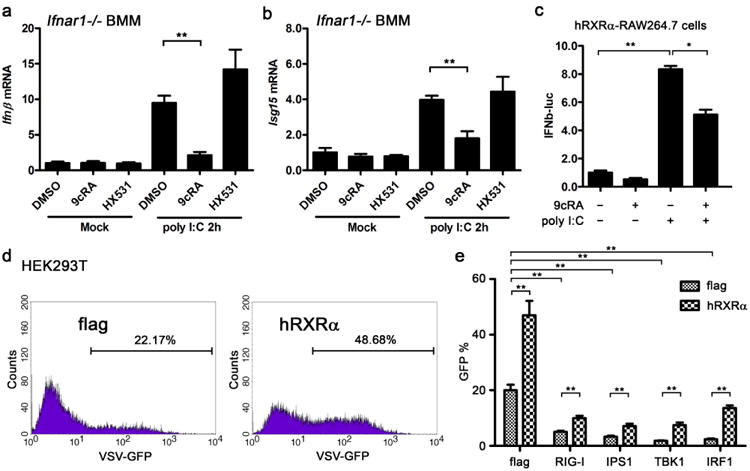
(a,b) Ifnar−/− BMMs were pretreated with DMSO, 9cRA (100 nM) or HX531 (1 μM) for 24 h and transfected with 1 μgml−1 polyI:C. Ifnβ and Isg15 mRNA were quantified by qPCR. Data are shown as mean ± s.d. (n = 3) of one representative experiment, similar results were obtained in three independent experiments, **P<0.01 (Student's t-test). (c) hRXRα-stably transfected RAW264.7 cells were transfected with IFNβ promoter luciferase reporter (IFNβ-luc) and treated with DMSO or 9cRA (100 nM) for 9 h before stimulation with polyI:C (5 μgml−1). The lysates were collected 20 h after polyI:C stimulation and luciferase reporter activity was measured by luminescence assay. RLU is represented as mean ± s.d. (n = 6) and is representative of three experiments. *P<0.05 and **P<0.01 (Student's t-test). (d) Flag or hRXRα vector was transfected into HEK293Tcells for 24 h, subsequently the transfected cells were infected by VSV-GFP (MOI = 0.01) for 8 h, fluorescence-activated cell sorting (FACS) assay was performed to detect the GFP-positive cells as VSV-infected cells. Data are representative of three independent experiments. (e) Flag or hRXRα was co-transfected with RIG-I and TLR3 pathway components for 24 h into 293T cells, subsequently the transfected cells were infected by VSV-GFP (MOI = 0.01) for 8 h, FACS assay was performed to detect the VSV virus in the cells. The ratio of GFP-positive cell is represented as mean ± s.d. (n = 3) and is representative of three experiments, P<0.01 (Student's t-test).