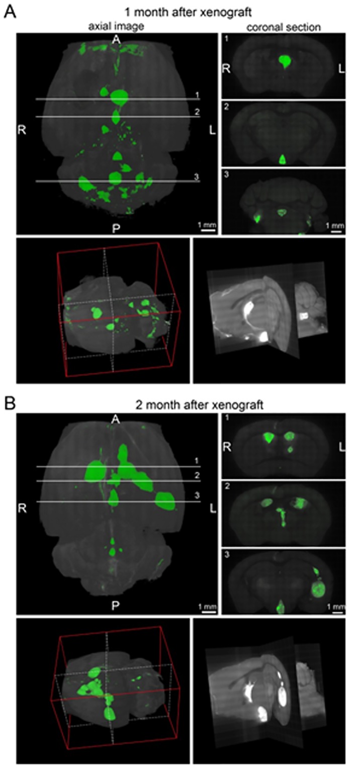
Fig 3. 3D imaging of the mice brain using a confocal microscope at one month A- or two months B- post—injection (LCAS-R).

Top left panels reconstitute images of the brains placed horizontally, top right panels reconstitute coronal sections at the indicated lines 1–3. Bottom left panels reconstitute 3D image of the brains oriented as shown by the red plot lines. The section images at the white dotted lines shown at the bottom right panels. A—anterior; P—posterior; R-right; L—left.
