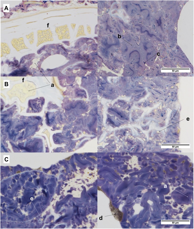
Fig 7. Micrographs showing the microstructure at the center of the crisp bread.
(A) unfermented whole grain rye crisp bread (uRCB), (B) yeast-fermented whole grain rye crisp bread (RCB), and (C) yeast-fermented refined wheat crisp bread (WCB). Protein is colored yellow (a), amylopectin-rich areas purple (b), and amylose blue (c). Fat can be seen in WCB as brown aggregates (d) and yeast cells are present in WCB and RCB (e). Aleurone layers can be seen in uRCB and RCB (f).

