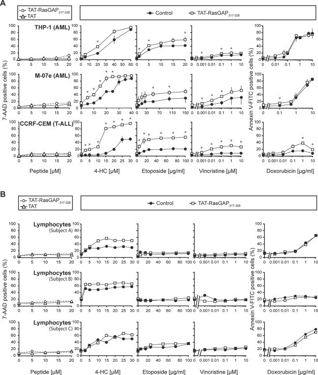
Fig 2. The effect of TAT-RasGAP317-326 as a chemosensitizer of leukemia cells and non-tumor lymphocytes.
A. Two acute myeloid leukemia cell lines (THP-1 and M-07e) and one T acute lymphoblastic leukemia cell line (CCRF-CEM) were seeded in 6-well plates and directly treated with 4-HC, etoposide, vincristine or doxorubicin at the indicated concentrations in the presence or in the absence of 10 μM TAT-RasGAP317-326. After 24 hours of drug incubation, 7-AAD or Annexin V-FITC staining was performed to evaluate cell death (last four columns). Alternatively (first column), the cells were treated with increasing concentrations of TAT or TAT-RasGAP317-326 alone. After 24 hours, the evaluation of cell death was carried out using 7-AAD staining. B. Isolated lymphocytes from three distinct healthy subjects were treated as described in Fig. 2A. The dosages of chemotherapeutic agents used to treat the healthy lymphocytes from the three subjects are similar to those used to treat the T-ALL CCRF-CEM cells. Note that the graphs are derived from single experiments where lymphocytes are immediately used after their isolation (if cultured in vitro, they would experience high levels of spontaneous apoptosis that would prevent accurate measurement of anti-cancer drug- and peptide-induced death). T-ALL, T-acute lymphoblastic leukemia; AML, acute myeloid leukemia; 4-HC, 4-hydroperoxycyclophosphamide. * p<0.05 t-test after Bonferroni correction.

