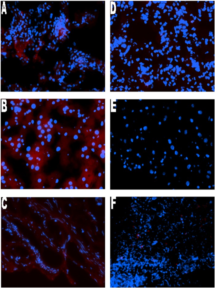
Fig 4. Immunofluorescence staining of rat tissue with villin.
Representative case of (A)metastatic lung, (B) metastatic liver, (C) metastatic stomach, (D)non-metastatic lung, (E) non-metastatic liver and (F) non-metastatic stomach. Positive villin cytoplasmic staining was detected in all metastatic samples with the Alexa Fluor 594 secondary antibody, conjugated to a red fluorophore. Metastasis negative liver, lung and stomach show the absence of villin staining.

