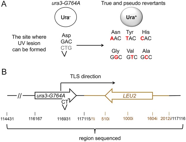
Fig 3. A genetic system to analyze the products of TLS through a chromosomal UV lesion.
(A) The ura3-G764A allele and possible UV-induced single nucleotide substitutions that lead to the Ura+ phenotype (marked in red). The sequence of the non-transcribed DNA strand is in black, and the transcribed strand is in grey. The site of potential UV lesion formation is indicated with a “V”. (B) A schematic showing the structure of the ura3-G764A-LEU2 cassette in chromosome V, the direction of the UV lesion bypass, and the region that was analyzed by DNA sequencing. The 2-kb HpaI LEU2 fragment used as a selectable marker for introducing the ura3-G764A allele into the chromosome is in dark yellow. Open arrows indicate open reading frames. Black numbers show chromosomal nucleotide position in respect to the left telomere; dark yellow numbers with the “i” index show nucleotide position within the LEU2 insert in respect to the end of the HpaI fragment.

