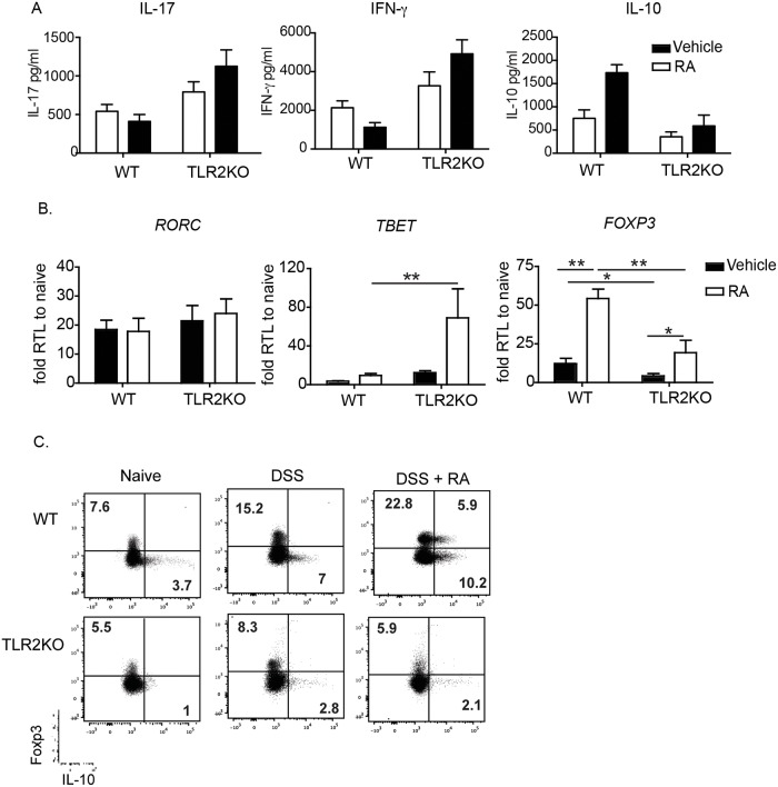
Fig 2. RA treatment suppresses IFN-γ during DSS in WT mice but enhances their secretion in TLR2KO mice.
(A) Mucosal scrapings from mice receiving the DSS and water were harvested on day 10 and analyzed by ELISA for cytokine levels. (B) Quantitative RT-PCR for transcription factors associated with T helper subsets were also performed on colonic lamina propria samples taken at day 14. Data shown are the fold increase of Vehicle- and RA-treated mice compared to naive controls. Data are the mean ± SEM of 5–8 mice per group pooled from two independent experiments. (C) Expression of Foxp3 and IL-10 in colonic LP CD4+ T cells (n = 3 per group), one representative facs plot is shown. *, p < 0.05, **, p < 0.01 using Student’s t-test.

