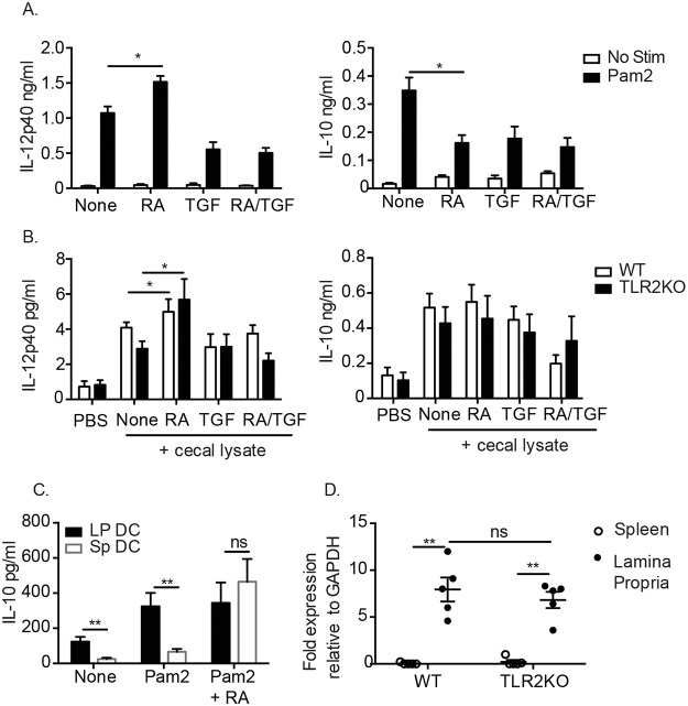
Fig 3. RA potentiates cytokine responses in both WT and TLR2KO DC.
(A) WT splenic DC were cultured with 100 ng/ml of TLR2 ligand Pam2CysK4 and (B) WT and TLR2KO splenic DC were cultured with 10 μg/ml cecal lysate in the presence or absence of RA, TGF- β or RA/TGF- β. Supernatants were analyzed after 24 hours for cytokine production by ELISA. (C) Comparison of IL-10 IL- production from CD103+ LP DC and CD103+ SP DC. (D) Transcript levels of aldh1a2 in spleen and LP of naïve WT and TLR2KO mice determined by quantitative PCR. For (A-C), data are the mean ± SEM of three independent experiments, for (D) 5 individual mice were analyzed. *, p < 0.05, **, p < 0.01 using Student’s t-test.

