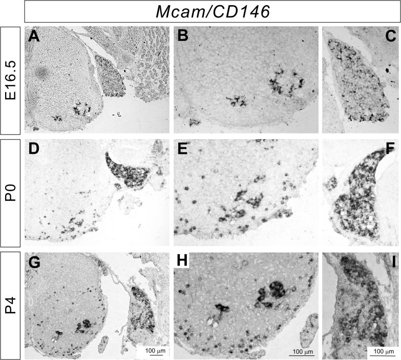
Fig 4. Expression of Mcam in the developing DRG and spinal cord.
(A-I) In situ hybridizations for Mcam on lumbar spinal cord sections from E16.5 (A-C), P0 (D-F), and P4 (G-I) wild-type mice. Mcam was strongly expressed by a subset of sensory and motor neurons at all stages. Expression of Mcam in the white matter was detected at P0 and P4 (D, E, G, and H).

