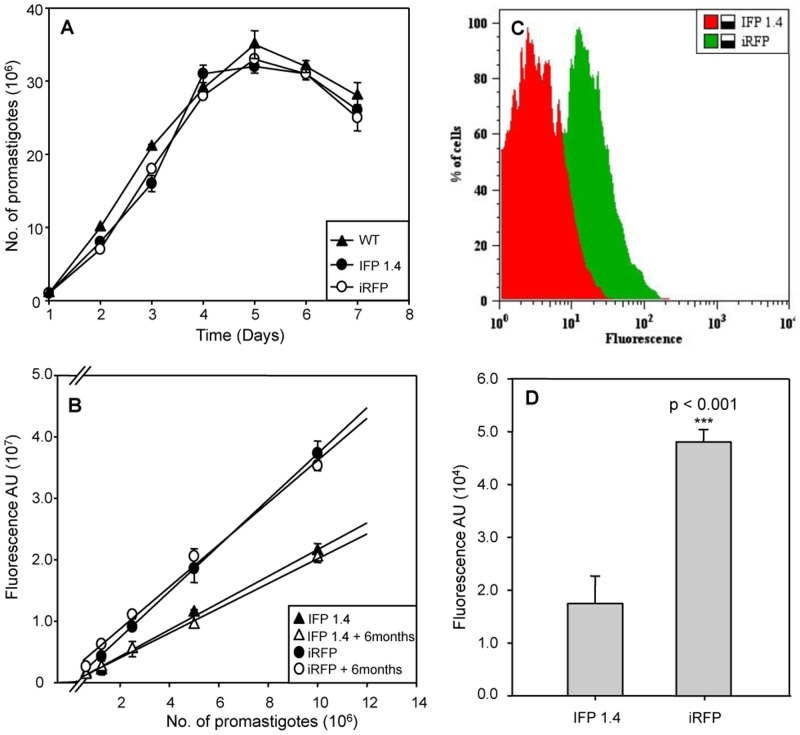
Fig 1. Infrared IFP1.4 and iRFP genes are functionally expressed in L. infantum parasites.
A) Growth rate of wild-type (WT; ▲) and stably-modified promastigotes IFP1.4+L.infantum (●) and iRFP+L.infantum (○). Parasites were counted using a Coulter counter. B) Correlation between fluorescence signal expressed as arbitrary units (AU) and the number of logarithmic IFP1.4+L. infantum (●) and iRFP+L. infantum (○) promastigotes in the presence of hygromycin B or after 6 months culture without drug IFP1.4+L. infantum (▲), iRFP+L. infantum (Δ). Two-fold serial dilutions were applied. C) Flow-cytometry analysis of intracellular amastigotes isolated from THP-1 in vitro infections with IFP1.4+L. infantum (red) and iRFP+L. infantum (green) strains. D) Mean fluorescence intensity emitted by lesion-derived amastigotes obtained from the engineered parasites. Experiments were carried out with 107 parasites by triplicate and error bars represent standard deviations. Significance level *** p< 0,001; ** p<0.01; from two tails of Student t-test.

