
Fig. 4.
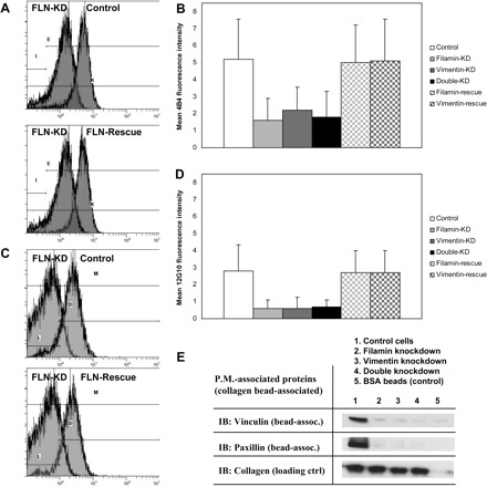
Filamin and vimentin regulate β1 integrin expression and activation. A: flow cytometry indicates decreased cell surface expression of β1 integrins following knockdown of filamin (FLN-KD, top). Cell surface levels of β1 integrin are restored by rescue of filamin expression (bottom). B: histogram shows mean 4B4 fluorescence intensity ± SD in control, filamin-KD, vimentin-KD, double-KD, filamin-rescue, and vimentin-rescue cells. C: flow cytometry indicates decreased β1 integrin activation following knockdown of filamin (top). Levels of β1 integrin activation are restored by rescue of filamin expression (bottom). D: histogram shows mean 12G10 fluorescence intensity ± SD in control, filamin-KD, vimentin-KD, double-KD, filamin-rescue, and vimentin-rescue cells. E: immunoblots (IB) of plasma-membrane (P.M.)-associated proteins show decreased recruitment of vinculin and paxillin to focal adhesions following knockdown of filamin and/or vimentin.