Fig. 6.

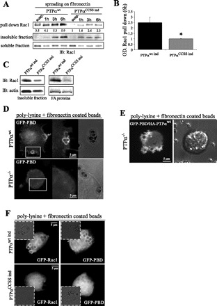
PTPα is required for Rac1 activation and localization at cell-matrix contacts. A: genetically modified NIH-3T3 cells expressing PTPαwt ind and PTPαCCSS ind were allowed to spread for 1, 3, and 6 h or kept in suspension (susp). Pull-down assay with glutathione S-transferase-tagged p21-activated kinase (PAK1)-binding domain (PBD) was used to purify active Rac1 from whole cell lysates, and Rac1 binding was analyzed by Western blotting. B: densitometry analysis of blot in A. OD, optical density. Values are means ± SD of 3 independent experiments. *P < 0.01 vs. PTPαwt ind (by unpaired Student's t-test). C: Western blot of Rac1 and actin in PTPαwt ind- and PTPαCCSS ind-expressing cells plated for 16 h. Triton X-100-insoluble fraction and FA-associated proteins are shown. D: images acquired from PTPαwt and PTPα−/− fibroblasts transiently transfected with GFP-tagged PBD and allowed to spread on fibronectin-coated beads for 30 min. E: images acquired from PTPα−/− cells cotransfected with GFP-PBD and HA-PTPαwt vectors and allowed to spread on fibronectin-coated beads for 30 min. F: images acquired from PTPαwt ind- and PTPαCCSS ind-expressing cells transfected with GFP-Rac1 or GFP-PBD and allowed to spread on fibronectin-coated beads for 30 min.
