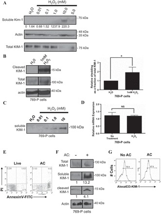
Fig. 1.

H2O2 and excess apoptotic cells accelerate Kidney injury molecule-1 (KIM-1) shedding. A: primary mouse proximal tubule epithelial cells (PTECs) were treated with increasing concentrations of H2O2, and both conditioned media and total cell lysates were collected. Soluble mouse KIM-1 was detected by SDS-PAGE and Western blotting. Soluble KIM-1 relative to total KIM-1 was quantified by densitometry. B: 769-P cells were exposed to H2O (1 μl) or H2O2 (1 mM) for 30 min in serum-free DMEM and the total cell lysates were collected and analyzed by Western blotting for cleaved and total KIM-1. The graph summarizes cleaved KIM-1 relative to total KIM-1 as determined by densitometry from 3 independent experiments (right). C: confluent monolayers of 769-P cells plated in 6-well tissue culture plates were exposed to H2O (1 μl) or various concentrations of H2O2 (0.01, 0.1, 1.0, and 10 mM) for 30 min in serum-free DMEM before analysis of the conditioned medium for soluble KIM-1. D: H2O2-induced KIM-1 shedding occurs independently of changes in KIM-1 mRNA expression. 769-P cells were exposed to H2O (1 μl) or H2O2 (1 mM) for 30 min in serum-free DMEM. Total cellular RNA was harvested, and relative KIM-1 mRNA expression was quantified using quantitative RT-PCR. E: UV-induced apoptotic(AC) or live mouse thymocytes that were stained with annexin V-FITC and propidium iodide (PI) and analyzed by flow cytometry. F: confluent monolayers of 769-P cells were not exposed or exposed to 107 apoptotic cells for 1 h. Relative shedding as a fraction of total KIM-1 was quantified by densitometry. A–E: soluble KIM-1 was detected from media samples with AKG antibody. Cleaved and total KIM-1 were detected in the total cellular lysates with 195 antibody. Actin was used as a loading control. The Western blot result is representative of 3 independent experiments. G: after being not fed or fed 5-(and 6)-carboxyfluorescein di-acetate succinimidyl ester (CFSE)-labeled apoptotic cells (AC) as in D, single-cell suspensions of the 769-P cells were analyzed for change in surface KIM-1 expression (Alexa 633-KIM-1) after staining with anti-KIM-1 primary antibody (AKG) and Alexa 633-conjugated secondary antibody. NS, not significant. *P < 0.05.
