
Fig. 2.
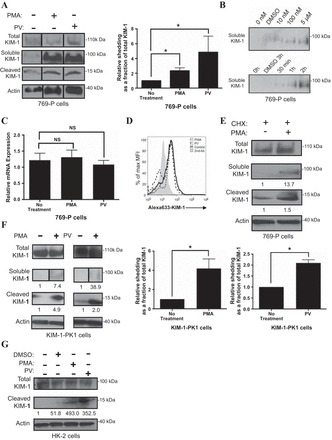
PMA and pervanadate (PV) accelerate KIM-1 shedding. A: confluent monolayers of 769-P cells were incubated in serum-free DMEM with DMSO (control) or PMA, (1 μM) for 1 h or PV (50 μM) for 30 min. A graph summarizing soluble KIM-1 relative to total KIM-1 in 769-P cells as determined by densitometry from 3 independent experiments is shown. B: 769-P cells were pretreated with PMA at various concentrations (0, 10, and 100 nM, and 5 μM) for 30 min (left) and for various durations of time (30 min, 1 h, or 2 h) at a concentration of 1 μM (right). C: PMA-induced KIM-1 shedding occurs independently of mRNA synthesis. 769-P cells were exposed to DMSO (control) or PMA for 1 h in serum-free DMEM. Total cellular RNA was harvested, and relative KIM-1 mRNA expression was analyzed using quantitative RT-PCR. D: 769-P cells were incubated for 30 min with or without PMA (1 μM) or PV (50 μM) before detection of surface KIM-1 expression by flow cytometry using AKG antibody and Alexa Fluoro 633-conjugated as secondary antibodies. KIM-1 expression is displayed in the form of a single-parameter (Alex 633-KIM-1) histogram against the % maximum mean fluorescent intensity. E: PMA-induced KIM-1 shedding occurs independently of protein synthesis. 769-P cells were preincubated for 30 min with cycloheximide (CHX; 25 μg/ml) in DMEM medium and then, with DMSO (control) or PMA (1 μM) for an additional 1 h before detection of soluble, cleaved, and total KIM-1 by Western blotting. F: KIM-1-PK1 cells were incubated in DMEM with DMSO (control) or PMA (1 μM) for 1 h, or with or without PV (50 μM) for 30 min. Graph summarizing cleaved KIM-1 relative total KIM-1 in KIM-1-PK-1 cells as determined by densitometry from 3 independent experiments is shown. G: HK-2 cells were incubated in DMEM with DMSO (control) or PMA (1 μM) for 1 h, or PV (50 μM) for 30 min. A–G: soluble KIM-1 was detected from media samples with AKG antibody. Cleaved and total KIM-1 were detected in the total cellular lysates with 195 antibody. Actin was used as a loading control. Cleaved KIM-1 relative to total KIM-1 was quantified by densitometry. Western blot results described above are representative of results obtained from 3 independent experiments. *P < 0.05.