
Fig. 4.
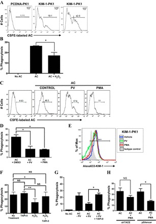
Accelerated KIM-1 shedding inhibits efferocytosis in KIM-1-expressing PTECs. A: PCDNA-PK1 or KIM-1-PK1 cells were incubated with 106 or 107 CFSE-labeled apoptotic cells(AC) in 6-well plates for 90 min, and the phagocytic uptake of apoptotic cells was analyzed by flow cytometry. Data re depicted in the form of a representative single-parameter (CFSE-labeled AC) histogram determined from 3 independent experiments. B: KIM-1-PK1 cells were untreated or pretreated with H2O (control) or H2O2 (1 mM) for 30 min in 6-well plates followed by incubation with 107 CFSE-labeled apoptotic cells for 90 min before measurement of phagocytic uptake of apoptotic cells by flow cytometry. C: KIM-1-PK1 cells were pretreated with DMSO (control), PV (50 μM) for 30 min, or PMA (1 μM) for 1 h in 6-well plates followed by incubation with 107 CFSE-labeled apoptotic cells for 90 min. Samples were analyzed by flow cytometry as in A. D: data from 3 independent experiments in C were graphed to show percent phagocytosis with error bars representing SD. E: change in surface KIM-1- expression in KIM-1-PK1 cells after addition of DMSO (vehicle), PMA, and PV as determined by flow cytometry using AKG primary antibody and Alexa Fluoro 633-conjugated anti-mouse IgG secondary antibody. F: 769-P cells were first pretreated with TAPI-0 (10 μM) for 30 min and then with H2O2 (1 mM) for 30 min before incubation with 106 CFSE-labeled apoptotic cells for an additional 90 min. Percent phagocytosis was determined as described in A from 3 independent experiments. G: KIM-1-PK1 cells were first pretreated with TAPI-0 (10 μM) for 30 min and then with DMSO (−PV) or PV (+PV) for another 30 min before incubation with 106 CFSE-labeled apoptotic cells for an additional 90 min. Percent phagocytosis was determined as described in A. H: shTACE and pSilencer cells were pretreated with and without PMA (1 μM) for 1 h followed by incubation with apoptotic cells for 90 min. Flow cytometry analysis was done as in A. *P < 0.05.