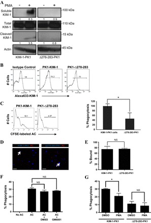
Fig. 6.

Phagocytic uptake of apoptotic cells is impaired in PTECs expressing a cleavage-mutant of KIM-1. A: LLC-PK1 cells stably expressing wild-type (KIM-1-PK1) or a cleavage-mutant of KIM-1 (Δ278–283-PK1) were treated with PMA (1 μM) or vehicle control (DMSO) for 30 min. Soluble and cleaved KIM-1 were detected from media and cell lysate samples, respectively. Actin was used as a loading control. Soluble and cleaved KIM-1 relative to total KIM-1 was quantified by densitometry. B: surface expression of KIM-1 in KIM-1-PK1 and Δ278–283-PK1 cells was evaluated by flow cytometry using both AKG or control antibody and Alexa Fluoro 633-conjugated secondary without permeabilization. C: KIM-1-PK1 and Δ278–283-PK1 cells were incubated with 106 CFSE-labeled apoptotic cells for 90 min, and phagocytosis was analyzed by flow cytometry. Data are depicted in the form of a representative single-parameter histogram (left) and a graph summarizing the relative (%) phagocytosis determined from 3 independent experiments (right). D: binding of pHrodo-labeled apoptotic cells(AC) to confluent monolayers of KIM-1-PK1 and Δ278–283-PK1 cells at 4°C as observed by confocal microscopy. Images were captured at 400× magnification, and the scale bar represents 10 μm. E: percent bound apoptotic cells relative to the total number of nuclei determined from 3 independent experiments in D. F: KIM-1-PK1 cells were pretreated with DMSO (control) or GM6001 (1 μM) for 30 min followed by incubation with 107 CFSE-labeled apoptotic cells (AC) for 90 min. Flow cytometric analysis was used to determine the percent phagocytosis from 3 independent experiments. G: KIM-1-PK1 and Δ278–283-PK1 cells were pretreated with DMSO (control) or PMA (1 μM) for 1 h in 6-well plates followed by incubation with 107 CFSE-labeled apoptotic cells for 90 min. Percent phagocytosis was determined by flow cytometry from 3 independent experiments. *P < 0.05.
