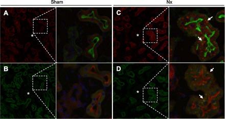
Fig. 5.

Immunofluorescent analysis of Cyclin B2 and Cdc2 in the kidney. The kidney was perfused, fixed, and then embedded. Sections (5 μm) were stained with a specific antibody for Cyclin B2 (A and C, red) or Cdc2 (B and D, green), phalloidin (A and C, green; B and D, red), and DAPI (blue). A and B: series of sections from sham-operated rat kidney. C and D: series of sections from Nx rat kidney at 2 wk after surgery. Arrows, aggregation of signals for Cyclin B2 or Cdc2; *, glomeruli. Magnification ×200 and ×400.
