
Figure 5.
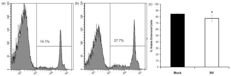
Assessment of cell viability in enteroids following exposure to rotavirus. Human jejunal enteroids were mock treated (a) or treated with rotavirus (b) for 24 h. Single cell suspensions were incubated with propidium iodide (PI) and 50,000 events examined for the presence of PI fluorescence using an LSRII flow cytometer. (a) and (b) Representative histograms showing percentage of cells that take up PI (non-viable cells) after each treatment. (c) Quantitation of viable cells after each treatment (n =3 ± SD). *P <0.05 using Mann–Whitney test